
Liver fibrosis in nonalcoholic fatty liver disease patients: noninvasive evaluation and correlation with cardiovascular disease and mortality

 , ...
, ... Abstract
Liver fibrosis is critical for liver-related outcomes and mortality in chronic liver disease, irrespective of etiology, including nonalcoholic fatty liver disease (NAFLD). NAFLD has been viewed as an independent correlate of cardiovascular risk. This review article briefly describes the cellular and molecular pathomechanisms underlying hepatic fibrosis. We then address noninvasive assessment of liver fibrosis. Finally, we discuss published evidence supporting fibrosis biomarkers’ role in assessing cardiovascular risk among patients with NAFLD. While histological assessment is the diagnostic standard of hepatic fibrosis, we specifically address noninvasive techniques, including equations based on anthropometric parameters, laboratory indices, and elastometry obtained with imaging techniques. The former group includes AST: ALT ratio, the Forns Index, the AST-to-platelet ratio index score, BARD (BMI, AAR, Diabetes) score, the fibrosis-4 index (FIB-4), the NAFLD fibrosis score, the gamma-glutamyl transferase-to-platelet ratio, and the Hepamet fibrosis score. The latter comprises elastographic techniques associated with ultrasonography or magnetic resonance. Our literature review identified numerous studies demonstrating that biomarkers of fibrosis (the most common being FIB-4) and elastographic techniques predict overall mortality and major cardiovascular events among NAFLD patients. The mechanisms accounting for this association are briefly reviewed. In addition to assessing hepatic fibrosis at baseline, during follow-up, and after therapeutic interventions in NAFLD patients, noninvasive assessment of hepatic fibrosis may predict cardiovascular events and overall mortality in these patients.

Keywords
INTRODUCTION
Liver fibrosis is associated with pathogenically diverse triggers and describes the excess accumulation of extracellular matrix (ECM) components that result from persistent liver injury[1,2]. Evolutionarily, fibrogenesis maintains tissue integrity by encapsulating an offending agent and limiting tissue damage; however, given sufficient time and whenever the fibrotic response exceeds a physiological amount, fibrosis impairs liver regeneration and jeopardizes hepatic function[3]. This notion accounts for increasing healthcare expenditures associated with advanced liver fibrosis stages[4].
Irrespective of the specific pathogenic type of inciting hepatic insult (e.g., viral, alcoholic, metabolic, autoimmune, cholestatic, drug-induced, or inherited), repeated bouts of hepatitis, hepatocyte damage, and wound-healing fibrosing response, if left untreated, result in progressive hepatic fibrosis and cirrhosis[5,6]. Cirrhosis is a fully de-structured liver histology characterized by accumulating large quantities of connective scar tissue and nodular architecture resulting in portal hypertension, progressive liver failure, or hepatocellular carcinoma[7]. Fibrosis is the sole histologic feature of nonalcoholic steatohepatitis (NASH) that predicts significant clinical outcomes such as cirrhosis, liver failure, and hepatocellular carcinoma[8-10]. Nevertheless, the major overall cause of mortality in patients with NASH is cardiovascular disease (CVD), especially in patients who do not yet have advanced cirrhosis, while significant hepatic-related morbidity and mortality are strictly associated with NASH-cirrhosis[11]. Seen from another perspective, but consistently with these notions, Mantovani et al. performed a meta-analysis of 5,802,226 adults in 36 published longitudinal studies over a median follow-up of 6.5 years and found that NAFLD was associated with a moderately increased risk of fatal or non-fatal CVD events[12]. However, this risk markedly increased across the severity of NAFLD, especially the fibrosis stage, irrespective of confounding metabolic factors[12].
While the diagnosis of cirrhosis may be clinically apparent in more advanced cases, earlier forms of the disease may require liver histology assessment[7]. Liver biopsy, however, is an invasive procedure with inherent sampling issues and observer variability and cannot be proposed other than for selected NAFLD patients, particularly in therapeutic trials[13,14]. On the one hand, hepatic fibrosis is a major determining factor of outcomes in chronic liver disease of all etiologies (NAFLD included); on the other hand, liver biopsy cannot be proposed extensively. The present study, therefore, focuses on the role of biomarkers and imaging techniques in diagnosing liver fibrosis and prognostication of the cardiovascular risk (CVR), which is inherent in advanced NAFLD forms.
CELLULAR AND MOLECULAR PATHOPHYSIOLOGY OF HEPATIC FIBROSIS
Mechanistically, fibrogenesis occurs when stressed or damaged hepatocytes release substances such as reactive oxygen species and activate macrophages and lymphocytes to generate several types of cytokines, including transforming growth factor-β and platelet-derived growth factor[15]. Collectively, these cytokines result in a net imbalance between increased ECM synthesis on the one hand and ECM degradation on the other, which results in hepatic fibrosis[15].
Several systemic signals from extra-hepatic tissues promote pro-fibrogenic responses in the liver, directly or indirectly acting on hepatic stellate cells (HSCs)[15]. For example, endotoxin, gut-derived pathogen-associated molecular patterns, and danger-associated molecular patterns are fibrogenic by signaling through TLR4 and other innate immune receptors[6]. This finding implies that the gut is a significant driver of NASH fibrosis through the microbiome’s effect and its interaction with the gut-liver axis, although the relative contributions of liver-derived and systemic molecules remain uncertain[6].
Morphologic patterns of fibrosis progression differ according to the etiology of liver disease, and, in NASH, fibrogenesis begins with the capillarization of sinusoids in zone 3 that progressively becomes panlobular (so-called “pericentral fibrosis”)[3]. NASH-specific fibrogenic pathways are increasingly being identified. Because metabolic stress induces the synthesis and release of pro-inflammatory chemokines, Kupffer cells gain inflammatory phenotypes contributing to the activation of HSC in concert with reactive oxygen species, apoptotic signals, and endoplasmic reticulum stress[16]. These pro-fibrogenic signals include oxidized phospholipids and enhanced signaling by the intra-hepatocytic transcriptional activator TAZ, which promotes paracrine fibrogenic signaling to HSCs via the secretion of Indian hedgehog ligand. The PNPLA3-I148M variant’s effects on HSC fibrogenesis increase ECM and collagen deposition, resulting in liver fibrosis[6].
Attesting to the biological complexity of fibrogenesis (which recruits different physiological pathways), the coagulation system is also involved, as initially reported by the pathologist Wanless (recently reviewed)[17-19]. This notion paves the way for anticoagulation strategies for treating NAFLD[18].
The cell type responsible for hepatic fibrogenesis is the peri-sinusoidal resident HSC, which is activated to gain a myofibroblast phenotype[6]. Therefore, all factors regulating the individual steps from HSC activation, proliferation, function, and survival represent essential therapeutic targets[15]. Additional therapeutic options include the elimination of the hepatotoxic agent(s) and (importantly) degradation of excessive ECM deposition[15]. Innovative approaches include manipulation of the coagulation cascade, anti-platelet drugs, and targeting integrins, which are master regulators of transforming growth factor-β activity[18,20-22]. Finally, cell therapy approaches are also under investigation[23].
Collectively, management approaches support the relatively recent notion that the complex fibrogenic process is not irreversible, offering a chance for the prevention and therapy of fibrosis[3,24,25].
The demonstration that, by treating liver fibrosis, we also address CVD would represent strong proof-of-concept evidence for a causal association between advanced liver disease and major cardiovascular events (MACE). However, rather than an improbable silver bullet, the reversal of liver fibrosis presently requests a holistic approach ranging from lifestyle changes to surgery[25,26]. Halfway between diet, exercise, and weight loss on the one end and bariatric surgery on the other end, there could be a pharmacological approach to treating fibrosis associated with NASH. This topic (beyond the scope of the present review) has been covered by Friedman et al.[6]. Among the various possible strategies, we highlight the study by Yang et al., who fed a choline-deficient amino acid diet to rats and found that histological features of NASH and fibrosis were significantly attenuated by treatment with 4-methylumbelliferone, an inhibitor of hyaluran synthesis
NONINVASIVE PREDICTORS OF LIVER FIBROSIS
Liver histology is the standard in evaluating liver fibrosis; however, this procedure has some limitations, such as invasiveness. Moreover, it carries the risk of complications. Finally, it is limited by sampling errors and intra- and inter-observer variability[14,31]. Owing to these drawbacks, research on diagnostic tools for accurately and noninvasively staging fibrosis is ongoing, and several fibrosis assessment tools have been developed to avoid liver biopsy in a subset of patients[32].
Given that imaging techniques tend to be available only among referral centers owing to substantial costs, general clinical scoring systems and combined assessment of laboratory biomarkers for liver fibrosis determination will be discussed first[32].
Serum liver biomarkers
Diagnosis of liver fibrosis with noninvasive biomarkers
The so-called “direct” fibrosis biomarkers (including ELF, FibroTest, FibroMeter NAFLD, Hepascore, ADAPT, FIBC3, ABC3D, and others) are not universally available. Moreover, some are patented and incur a fee. These tests are discussed in detail elsewhere and are beyond the scope of this review[33-35].
Several “indirect” biomarkers of fibrosis, based on a combination of anthropometric and serum variables, are freely available globally for clinical practice. They are listed in Table 1, which also illustrates the diagnostic accuracies[36-44].
Indirect biomarkers of liver fibrosis
| Biomarker | Author (ref.) | Equation | Cut-offs for F3/F4 | AUROC* |
| AST: ALT ratio (AAR) | Williams et al.[36] | AST (IU/L)/ALT (IU/l) | < 0.8 to rule out > 1.0 to rule in** | - |
| Forns index | Forns | 7.811-3.131 ln [platelet count (×109/L)] + 0.781 ln [GGT (IU/L)] + 3.467 ln [age (years)]-0.014 [cholesterol (mg/dl)] | < 4.2 to rule out > 6.9 to rule in¶ | 0.81 |
| AST-to-platelet ratio index (APRI) score | Wai | AST (IU/L)/(ULN)/platelet count (×109/L) × 100 | ≤ 0.5 to rule out > 1.5 to rule in¶ | 0.88 |
| BARD score | Harrison et al.[39] | Weighted sum of BMI ≥ 28 kg/m2, 1 point AST/ALT ratio ≥ 0.8, 2 points Diabetes present, 1 point | ≥ 2 points to rule in | 0.81 |
| Fibrosis-4 index (FIB-4) | Sterling | Age × AST (IU/L)/platelet count (×109/L) × √ALT (IU/l) | < 1.45 to rule out > 3.25 to rule in | 0.765 |
| NAFLD fibrosis score (NFS) | Angulo | -1.675 + 0.037 × age (years) + 0.094 × BMI (Kg/m2) + 1.13 × IFG or T2D (yes = 1; no = 0 =) + 0.99 × AST/ALT ratio - 0.013 × platelet count (×109/L)-0.66 × albumin (g/dL) | < -1.45 (< 65 years) < 0.12 (> 65 years) to rule out^ ≥ 0.67 to rule in | 0.82 |
| GGT-to-platelet ratio (GPR) | Lemoine et al.[43] | GGT/ULN of GGT/platelet count (109/L) ×100 | > 0.32 to rule in | 0.76-0.93° |
| Hepamet fibrosis score (HFS) | Ampuero et al.[44] | 1/[1 + e (5.390-0.986 (if Age 45-64 years) - 1.719 (if Age ≥ 65 years) + 0.875 (if Male sex) - 0.896 (if AST 35-69 IU/L) - 2.126 (if AST ≥ 70 IU/L) - 0.027 (if Albumin 4-4.49 g/dL) - 0.897 (if Albumin < 4 g/dL) - 0.899 (if HOMA 2-3.99 with no DM) -1.497 (if HOMA ≥ 4 with no DM) - 2.184 (if DM) - 0.882 (if Platelets 155-219 × 1000/µL) - 2.233 (if Platelets < 155 × 1000/µL))] | < 0.12 to rule out > 0.47 to rule in | 0.84 |
Ballestri et al. evaluated various liver fibrosis biomarkers to identify significant/advanced fibrosis[AST: ALT ratio (AAR); AST-to-platelet ratio Index (APRI), fibrosis-4 (FIB-4) index, Forns index, BMI, AAR, Diabetes (BARD) score, and Hepamet fibrosis score (HFS)] in their series of 157 hepatitis C virus (HCV), 16 hepatitis B virus (HBV), and 107 NAFLD consecutive patients[45]. These individuals used the following scoring systems to estimate CVR: SCORE, Progetto CUORE, or the Framingham risk scoring systems. Data showed that biomarkers of hepatic fibrosis had a higher diagnostic performance in patients with advanced fibrosis (compared to those with significant fibrosis). The Forns Index and HFS showed the best performance for NAFLD in identifying advanced fibrosis. By lowering their cut-off values, these fibrosis biomarkers exhibited high negative predictive values (NPVs) for advanced-stage fibrosis. Interestingly, FIB-4, Forns index, NFS, and HFS were positively correlated with SCORE and Framingham risk scores. Collectively, these findings support the notion that biomarkers of liver fibrosis can exclude advanced fibrosis while positively correlating with CVR scores[45]. A more recent study by Brandman et al., conducted on 1483 individuals with biopsy-proven NAFLD recruited in the USA or the UK, found that two scores most accurately identified cirrhotic patients: FIB-4 and NFS[area under the receiver operating characteristic curve (AUROC) 0.84-0.86)][46].
It remains uncertain which of the various noninvasive fibrosis tests is best in clinical practice, and the choice depends on the cut-off used. Confirming this notion, Sun et al. performed a meta-analysis of four studies with 1038 adult patients and found that FIB-4 at a cut-off threshold of 1.30 was more accurate than a cut-off of 3.25, NFS and BARD; however, FIB-4 has a limited capacity to predict advanced fibrosis in patients with NAFLD[47]. The limitations inherent in these noninvasive fibrosis biomarkers should not be neglected. For example, Boursier et al. evaluated 1051 biopsy-proven NAFLD patients in whom serum blood fibrosis markers (NFS; FIB4; Fibrotest; FibroMeter), vibration-controlled transient elastography (VCTE), and the combinatory elasto-blood test FibroMeterVCTE. The authors found that the AUROCs were significantly lower among type 2 diabetes (T2D) patients, mainly owing to decreased specificity. The reasons underlying this finding are that these noninvasive tests are biased by the different characteristics of the groups studied but are linked to T2D per se, which carries modifications of the levels of some noninvasive biomarkers of fibrosis[48].
Collectively, these studies support the notion that combining serum biomarkers and biomarkers based on imaging techniques offers increased accuracy in the noninvasive assessment of significant hepatic fibrosis among those with NASH. Whether such combined indices also predict CVR more accurately than currently available biomarkers and techniques remains to be ascertained.
Prediction of CVD and mortality
A consistent body of research has documented that biopsy-proven liver fibrosis is robustly associated with overall and liver-related mortality[55]. Noninvasive fibrosis scores, including AAR, FIB-4, and NFS, identify individuals prone to the risk of progressive fibrosis[56]. Therefore, the logical question is whether (and to what extent) the same predictive power is retained by noninvasive biomarkers.
In their seminal study, Perazzo et al. prospectively followed 2312 patients with T2D or dyslipidemia for 5-15 years and found that advanced fibrosis, defined by a FibroTest > 0.48, were significantly associated with overall mortality[hazard ratio (HR) 1.95, 95% confidence interval (CI) 1.12-3.41], advancing to fibrosis at baseline (HR 0.92, CI: 1.04-3.55), and worsening to advanced fibrosis over time (HR 4.8, CI: 1.5-14.9)[57]. Moreover, among patients with an increased CVR (defined by a Framingham risk score ≥ 20%), advanced fibrosis predicted cardiovascular events (HR 2.24, CI: 1.16-4.33)[57]. After this pioneering study, many others confirmed that indirect biomarkers of fibrosis are associated with cardiovascular events and mortality, as summarized in Table 2[45].
Principal recent studies show the association between fibrosis biomarkers and indirect and direct evidence of increased CVD risk and mortality among NAFLD patients
| Author (ref.) | Series, method | Findings | Comment |
| Cross-sectional | |||
| Kim et al.[58] | 11,154 participants in whom NAFLD was defined using US evidence of steatosis in the absence of competing liver diseases were followed for a median of 14.5 years Liver fibrosis presence and severity were evaluated through NFS, APRI, and FIB-4 | 3.2% of participants had NFS > 0.676, indicative of advanced fibrosis The presence of NAFLD was not a risk factor for increased mortality. Conversely, mortality increased paralleling advancing fibrosis scores such that those at a high probability of advanced fibrosis had a 69% increase in mortality (for NFS: HR 1.69; for APRI: HR 1.85; for FIB-4: HR 1.66) vs. controls without fibrosis, after adjustment for age, sex, race-ethnicity, education, income, diabetes, hypertension, history of CVD, lipid-lowering medication, smoking status, waist circumference, alcohol, caffeine consumption, total cholesterol, HDL-C, transferrin saturation, and CRP Such increases in mortality were almost entirely accounted for by CVD (for NFS: HR 3.46; for APRI: HR 2.53; for FIB-4: HR 2.68) | While US-diagnosed NAFLD is not associated with increased mortality, advanced fibrosis, assessed with noninvasive fibrosis markers, significantly predicts mortality, mainly owing to CVD, independent of confounders |
| Song | 665 NAFLD subjects free of heart disease were enrolled NFS, FIB-4 score, Forn’s index, and the APRI were used to evaluate the severity of hepatic fibrosis | MVA identified age, BMI, and eGFR as significantly predicted CACS > 100 NFS, FIB-4 score, and APRI were significantly associated with a CAC score > 100 after adjusting: for age and sex (model 1) and age, sex, BMI, and eGFR (model 2) Predictive accuracy could be improved with integrated scores combining noninvasive biomarkers of hepatic fibrosis with other parameters | Widely used blood biomarkers of liver fibrosis can identify NAFLD patients with high CVD risk |
| Ballestri | 157 HCV, 16 HBV, and 107 NAFLD consecutive patients were enrolled The following liver fibrosis biomarkers were employed for capturing significant/advanced hepatic fibrosis: ARR; APRI; FIB-4; Forns index; BARD; and HFS CVR was assessed using the following scoring systems: SCORE, Progetto CUORE, or the FR scoring systems | Regardless of liver disease etiology, biomarkers of hepatic fibrosis performed better in predicting advanced rather than significant liver fibrosis In NAFLD patients: Forns index and HFS performed best in predicting advanced fibrosis Lower cut-offs of hepatic fibrosis biomarkers had high NPVs for advanced fibrosis FIB-4, Forns index, NFS, and HFS were positively correlated with SCORE and FR risk scores | In NAFLD (and chronic viral hepatitis), biomarkers of hepatic fibrosis can exclude advanced fibrosis and are associated with CVR scores |
| Schonmann | Historical data on 8511 individuals, 3292 with inconclusive fibrosis and 195 with advanced fibrosis (FIB-4 ≥ 2.67), were retrieved from a computerized medical database Hepatic fibrosis was assessed with the FIB-4 score, and the association with CVD was adjusted for SCORE | Individuals with advanced fibrosis had higher CVD risk after adjustment for sociodemographic characteristics, the SCORE, use of statins and aspirin (HR 1.63). The association persisted in both females and males When age-specific cut-offs were utilized, a significant dose-response association between inconclusive and advanced fibrosis and CVD was found (HR 1.15 and 1.60, respectively) | Fib-4, being independently associated with CVD, offers the opportunity for using markers of liver fibrosis in CVD risk assessment in primary care |
| Han | In 9444 KNHNES participants, liver fibrosis was assessed with FIB-4 and NFS A high ASCVD risk was defined as a 10-year ASCVD risk score > 10% | The quartiles of ASCVD risk scores were positively correlated to MAFLD and FIB-4 defined-significant liver fibrosis (P for trend < 0.001) Compared to MAFLD-free controls, individuals with both MAFLD and FIB-4 defined-significant liver fibrosis had a greater ASCVD risk (OR = 2.40; P < 0.001) (adjusted for sex, age, exercise, smoking, alcohol consumption, ASCVD history, SBP, fasting blood glucose, BMI, HOMA-IR, CKD, LDL-C, and muscle mass) The impact of MAFLD on a high probability of ASCVD risk was greater than that of significant liver fibrosis (OR = 4.72 for MAFLD vs. OR = 1.88 for FIB-4 defined-significant liver fibrosis; all P < 0.001) (adjusted for sex, age, exercise, current smoking, alcohol consumption, ASCVD history, CKD, LDL-C, and muscle mass) Among MAFLD individuals, having a low muscle mass was associated with a higher risk of significant liver fibrosis (OR = 1.56 to 2.43; P < 0.001), and similar findings were observed when significant hepatic fibrosis was defined with NFS | MAFLD per se carries a substantial ASCVD risk, further worsened by concomitant significant liver fibrosis. This, in turn, is associated with sarcopenia |
| Longitudinal | |||
| Chen | A prospective cohort study enrolling 3263 CAD Chinese patients submitted to a median follow-up period of 7.56 years (inter-quartile range: 6.86-8.31) Cox models were used to assess the association of baseline levels of NFS; FIB-4; APRI; GPR, and Forns score, with the risk of all-cause and CV mortality among CAD patients | 319 out of 538 deaths observed during this study were due to CVD Multivariable-adjusted HRs (95%CI) for those with the highest levels of NFS, FIB-4, APRI, GPR, and Forns score were 2.89 (2.14-3.91), 2.84 (2.14-3.76), 1.77 (1.33-2.36), 1.47 (1.19-1.83) and 3.10 (1.88-5.11) for all-cause mortality, 3.02 (2.05-4.45), 3.34 (2.29-4.86), 1.99 (1.40-2.83), 1.80 (1.36-2.39) and 2.43 (1.28-4.61) for CV mortality, respectively, compared to patients with lowest score levels These associations were consistent after ruling out early deaths (i.e., those occurring within the first year of follow-up) or stratifying patients (by sex, age, BMI, diabetes status, MetS status, CAD type, and hsCRP level) | Among CAD patients, higher liver fibrosis scores and noninvasive biomarkers are associated with increased mortality owing to all causes and CVD |
| Barattta | 898 consecutive outpatients (mean age, 56.4 ± 12.7 years; 37.5% women) underwent US to identify liver steatosis Liver fibrosis was defined as a FIB-4 score > 2.67 and NFS > 0.676 Phone interviews were conducted every 6 months, and patients were examined every 12 months in the outpatient clinic. Median follow-up time of 41.4 months (3044.4 patient-years) Primary outcomes: incidence rate of CVEs defined as fatal or nonfatal ischemic stroke and MI, cardiac or peripheral revascularization, new-onset AF, and CV death | CVEs rate was higher in patients with (2.1%/year) than without NAFLD (1.0%/year) (P > 0.05) At Cox MVA, NAFLD significantly increased the risk of CVEs (HR 2.41) after adjusting for MetS. Among NAFLD patients, male sex, previous CVEs, MetS, and FIB-4 scores > 2.67 (HR 4.02; P < 0.05) were independently associated with the risk of incident CVEs. NFS scores > 0.676 were also independently associated with the risk of incident CVEs (HR 2.35; P < 0.05) | Liver fibrosis indexes are strongly associated with incident CVEs among NAFLD individuals |
| Lee | 1173 asymptomatic adults with CAC scores from 2007-2013 were enrolled. Median follow-up: 3.0 (2.0-3.8) years CAC progression was defined as newly incident CAC or a ≥ 2.5-unit increase in the final CAC score square root Liver fibrosis was assessed with FIB-4 and NFS | The mean baseline FIB-4 score was significantly higher in subjects with CAC CAC progression rates were 20.5%, 27.5%, and 35.9% among those NAFLD-free, with NAFLD and low FIB-4 values, and those with NAFLD and intermediate/high FIB-4 scores, respectively At MVA, compared to NAFLD-free controls, subjects with NAFLD plus intermediate/high FIB-4 scores had OR for CAC progression = 1.70 after adjustment for sex, BMI, smoking, drinking, exercise, hypertension, T2D, serum TG, HDL-C, LDL-C, and hsCRP In the sensitivity analysis, the OR for CAC progression was 1.57 for subjects with NAFLD plus an intermediate/high NFS versus those without NAFLD | Noninvasively assessed advanced liver fibrosis associates with an increased chance of CAC progression among NAFLD patients |
| Liu | Multicenter, prospective study, enrolling 4003 consecutive patients with stable CAD undergoing PCI and followed up for an average of 5.0 ± 1.6 years the occurrence of MACE (CV death, non-fatal MI, and stroke) | Subjects who developed MACE were likelier to have either intermediate or high values of NFS; FIB-4; BARD; and ARR Moreover, compared to individuals who had low fibrosis markers scores, those with intermediate plus high score levels had significantly increased risk of MACE (aHR 1.57-1.92) after adjustment for age, sex, smoking, drinking, T2D, hypertension, SBP, LDL-C, HbA1c, hs-CRP, number of lesion vessels and baseline statin use Finally, the prediction ability of a model with established CV risk factors significantly improved by adding NFS, FIB-4, or BAARD | High values of hepatic fibrosis markers predict adverse outcomes in patients with stable CAD submitted to PCI, suggesting that these markers might also be evaluated in the risk stratification before elective PCI |
| Peters | Data from 1423 individuals with HFpEF from the TOPCAT trial were analyzed The risk of advanced liver fibrosis was classified as low, intermediate, and high based on NFS and FIB-4 scores Combined primary endpoints: all CV death, aborted cardiac arrest, and hospitalization for HF Cox MVA was used to test the association between the risk of fibrosis severity and the combined primary endpoint | Advanced fibrosis (defined by high fibrosis scores) was present in 37.57% of the NFS and 8.02% by the FIB-4 In unadjusted models, the risk of advanced fibrosis was significantly associated with the primary CV outcome (NFS high vs. low: HR 1.71; FIB-4 high vs. low: HR 1.56). However, this association was diminished (NFS high vs. low: HR 1.35, P > 0.05); FIB-4 high vs. low: HR 1.42, P = 0.05) after multivariable adjustment (sex, race, NYHA class, smoking, SBP, serum sodium, blood urea nitrogen, prior CVD, previous hospitalization for HF and ongoing spironolactone) | Advanced liver fibrosis (assessed with fibrosis biomarkers) may not be uncommon among HFpEF patients There seems to be a limited independent association between liver fibrosis risk scores and clinical outcomes related to HF events |
| Parikh | Nested case-cohort study within the REGARDS cohort: black and white participants aged ≥ 45 years recruited between 2003 and 2007 and followed up for ischemic stroke (median duration 5.4 years) FIB-4 and NFS were calculated using baseline data for stroke cases and a random cohort sample Cox proportional hazards models were used to estimate the HR of stroke after adjusting for potential confounders. Sex differences were assessed | 572 incident ischemic strokes were registered during follow-up. No significant association was found between liver fibrosis and ischemic stroke globally with FIB-4 and NFS. Nevertheless, such an association was found in women alone using FIB-4 | In women (but not men), there may be an association between advanced hepatic fibrosis and enhanced ischemic stroke risk |
| Oh | 4163 subjects from the Korean Genome and Epidemiology Study were followed biannually over a 15.6 median period Cox proportional hazards regression was used to gauge the HRs of either NAFLD or indexes of hepatic fibrosis in the global patient population and subsets identified with BMI and glucometabolic status | During follow-up, 643 subjects (15.4%) died. FIB-4, NFS, and APRI were consistently higher in deceased subjects regardless of baseline glucose metabolism status FIB-4 and NFS exhibited acceptable discrimination power for mortality, with AUROC values of 0.686 and 0.666, respectively. HRs for FIB-4 and NFS were 1.41 and 1.43, respectively (adjusted for age, sex, smoking, BMI, and HbA1c) FIB-4 and NFS were significantly associated with liver-specific mortality but not CV mortality The association between mortality with fibrosis indices was more marked among subjects with BMI < 25 kg/m2 | In Koreans, noninvasive assessment of fibrosis correlates with overall and hepato-specific mortality |
| Delgado | The impact of NAFLD noninvasive fibrosis indices was evaluated in a large cohort of German patients (3316 subjects) referred to coronary angiography between 1997 and 2000. Median follow-up of 9.8 years (range 0.1-11.3) | Modeling the common NAFLD and fibrosis scores, FIB-4 and NFS, as splines revealed significant associations with all-cause and CV mortality when Cox regression models were only adjusted for CVR factors that were not already included in the calculation of the scores Stratifying the scores into quartiles yielded HRs for all-cause and CV mortality for the fourth quartile versus the first quartile of 2.28 and 2.11 for FIB-4 and 3.21 and 3.12 for NFS However, an independent association of FIB-4 or NFS with overall or CV mortality was not observed in the prospective CAD cohort after full adjustment for age, sex, BMI, T2D, hypertension, smoking, LDL-C, HDL-C, medication, and alcohol intake | Noninvasive indices of fibrosing NAFLD do not offer additional data in assessing CVD events among angiography-proven CAD |
| Tamaki | 3512 subjects were enrolled in this prospective study between April 2019 and November 2020 Liver fibrosis was assessed noninvasively with FIB-4, NFS, and WFA+ -M2BP, and fatty liver was diagnosed with US. FRS ≥ 20% identified an elevated risk of CVD | FRS ≥ 20% was found in 17.5% Advanced fibrosis (FIB-4 ≥ 2.67, NFS ≥ 0.675, and WFA+ -M2BP ≥ 1.0) and the presence of fatty liver was significantly associated with high CVD risk independent of metabolic confounding factors (T2D, dyslipidemia, and hypertension) (OR 2.53-9.62) Advanced fibrosis and steatosis were associated with the most elevated CVD risk. Next were individuals with advanced hepatic fibrosis in the absence of steatosis | CVD risk was associated with liver fibrosis and steatosis irrespective of CVR copathologies suggesting that these hepatic conditions may help disclose individuals at a high CVR |
| Akuta | Retrospective investigation of the incidence and noninvasive predictors of CVDs, extra-hepatic cancer, and liver-related complications in 477 Japanese patients with biopsy-proven NAFLD followed for a median of 5.9 years | MVA FIB-4 ≥ 2.7 strongly predicted the three disease outcomes, independently of CKD, PNPLA3 rs738409, BMI, hypertension, and extra-hepatic malignancies Moreover, the global occurrence of CVDs significantly differed among the various PNPLA3 polymorphisms. These CVD risk factors could be attributed to CC PNPLA3 genotype, CKD, and FIB-4 ≥ 2.7 | Data have shown that the FIB-4 index ≥ 2.67 (and PNPLA3 genotype) affects the incidence of complications, particularly CVDs, among Japanese NAFLD patients |
| Jin | This study enrolled 5143 individuals with stable CAD demonstrated angiographically who were followed up for seven years CAD severity was assessed with the number of diseased vessels, Gensini, Syntax, and Jeopardy scores Outcome: predictive values of NFS and FIB-4 for CAD severity, CAC, and CVEs | Both NFS and FIB-4 predicted CAC. All CAD severity parameters were significantly more elevated among those with higher NFS scores At Kaplan-Meier analysis, patients with intermediate and high NFS and FIB-4 scores had a higher risk of CVEs and CV mortality At Cox MVA, NFS and FIB-4 were independently associated with CVEs [HR (95%CI): 1.150 (1.063-1.244), P < 0.001 and 1.128 (1.026-1.240), P = 0.012] (adjusted for sex, BMI, current smoking, T2D, hypertension, family history of CAD, left ventricular ejection fraction, creatinine, TG, LDL-C, HDL-C and baseline statin use) | NFS and FIB-4 were significantly associated with CAD severity, CAC, and CVEs |
| Zupo | 1929 elderly patients were recruited in a population study in southern Italy, followed for eight years Based on the FIB-4 score, patients were assigned to three liver fibrosis risk groups (low, intermediate, and high) For comparison purposes, the APRI score was also calculated in secondary analyses | The eight-year risk of death was almost two-fold among individuals in the highest-risk FIB-4 score group, even after controlling for possible confounding factors (age, sex, smoking, education, alcohol consumption, and multimorbidity) FIB-4 scores were associated with a steeper mortality curve than the APRI scores | In clinical practice, FIB-4 may help to identify individuals at risk of higher mortality |
| Zupo | 1929 elderly individuals Cardiovascular Health Study criteria were employed to classify physical frailty Mean observation time from a maximum of 56.87 ± 22.41 to a minimum of 48.49 ± 18.69 months Physical frailty + FIB-4 above 2.67 is defined as “liver frailty” Physical frailty, high-risk liver fibrosis, and liver frailty subjects were compared to non-frail controls Proportional Cox regression tested each category’s adjusted association between liver frailty and all-cause mortality | Compared to non-frail elderly individuals, liver frailty subjects were significantly older, with lower education and higher multimorbidity At Cox MVA, a two-fold increased overall mortality risk (HR 2.09) was found after adjusting for confounding factors (age, sex, education, and alcohol consumption) | Compared to non-frail controls, the liver frailty phenotype - defined based on simple measures primarily available in a primary care setting - is associated with a two-fold increased risk of overall mortality in a patient population from southern Italy |
| Vieira Barbosa | 81,108 patients with (a) NAFLD, (b) NASH, or (c) at risk of NASH. Median follow-up: 3 years Outcome: MACE (i.e., MI, hospitalization for unstable angina or HF, and coronary revascularization) | After adjusting for established CVR factors, FIB-4 ≥ 2.67 remained the strongest independent predictor of MACE overall (aHR 1.80) and was significantly associated with MI (aHR 1.46), hospitalization for unstable angina (aHR 1.24), hospitalization for HF (aHR 2.09), CABG (aHR 1.65), and PCI (aHR 1.72) | FIB-4 ranks as the most accurate predictive factor of MACE, irrespective of traditional CVR factors and hepatic diagnosis at the baseline |
Sonoelastography techniques
Diagnosis of liver fibrosis
Several ultrasound-based elastographic techniques have been implemented for the noninvasive assessment of liver fibrosis, including VCTE and shear wave elastography (SWE) techniques, divided into point-SWE (pSWE) such as acoustic radiation force impulse (ARFI) elastography and two-dimensional-SWE (2D-SWE) such as supersonic shear imaging (SSI)[76,77].
VCTE is the most widely used technique for the noninvasive staging of liver fibrosis in chronic liver disease and is evaluated using a dedicated tool called FibroScan[78]. VCTE uses a single hand-held probe that measures the velocity of low-amplitude shear waves by ultrasound, providing a point-of-care LSM. VCTE does not produce real-time sonographic images of the liver; however, the device is also equipped with a controlled attenuation parameter (CAP), a sensitive noninvasive method for diagnosing hepatic steatosis. Fibroscan enables simultaneous quantification of fibrosis and steatosis. To exclude significant/advanced fibrosis, an optimal cut-off threshold of 8 kPa has been proposed for the sequential use of noninvasive tests for risk stratification in NAFLD patients[78].
Conversely, ARFI and SSI are fully integrated into conventional ultrasound machines and, therefore, can be performed during routine abdominal ultrasound examinations. SWE techniques to generate shear waves utilize ultrasound impulses of high frequency; the operator is required to identify a region of interest and acquire a series of LSMs[79]. ARFI and SSI have shown reliable intra- and inter-observer measurement agreements[80]. Moreover, ARFI and SSI can selectively analyze specific liver points, including heterogeneous liver fibrosis or focal liver lesions[81].
VCTE, ARFI, and SSI have shown good accuracy for noninvasively assessing the degree of fibrosis in patients with biopsy-proven NAFLD, although fewer studies are available for SSI, and more data are requested regarding the best cut-off points for ARFI and SSI[52,82-85]. However, VCTE and SWE techniques are more accurate for detecting advanced fibrosis/cirrhosis (F3-F4 stages) than for mild-moderate (F1-F2) fibrosis[77,86]. The diagnostic performance of various elastographic techniques for detecting liver fibrosis stages (considering hepatic histology as the reference) among NAFLD individuals as reported in the most recent and most significant meta-analysis of 70 studies[53 VCTE, 11 pSWE, four 2DSWE, 11 MRE] are shown in Table 3[87]. According to this meta-analysis, the diagnostic performance of SSI was worse than reported in a previous meta-analysis of individual patient data, which were not included in the present meta-analysis[85].
Performance of elastographic techniques for detecting liver fibrosis stages in NAFLD (liver histology is used as the diagnostic standard)
| Imaging tool | Fibrosis severity | SE (%) | SP (%) | AUROC (95%CI) |
| VCTE (kPa) | F ≥ F2 | 80 | 73 | 0.83 (0.80-0.87) |
| F ≥ F3 | 80 | 77 | 0.85 (0.83-0.87) | |
| F = F4 | 76 | 88 | 0.89 (0.84-0.93) | |
| pSWE (m/s) | F ≥ F2 | 69 | 85 | 0.86 (0.78-0.90) |
| F ≥ F3 | 80 | 86 | 0.89 (0.83-0.95) | |
| F = F4 | 76 | 88 | 0.90 (0.82-0.95) | |
| 2DSWE (kPa) | F ≥ F2 | 71 | 67 | 0.75 (0.58-0.87) |
| F ≥ F3 | 72 | 72 | 0.72 (0.60-0.84) | |
| F = F4 | 78 | 84 | 0.88 (0.81-0.91) | |
| MRE (kPa) | F ≥ F2 | 78 | 89 | 0.91 (0.80-0.97) |
| F ≥ F3 | 83 | 89 | 0.92 (0.88-0.95) | |
| F = F4 | 81 | 90 | 0.90 (0.81-0.95) |
VCTE can also be used in clinical practice in cirrhotic patients to rule out significant portal hypertension with esophageal varices needing treatment, thus sparing unnecessary screening endoscopy examinations (e.g., NAFLD criteria: LSM < 30/25 kPa for the M/XL probe and platelet count > 110.000 mm3)[88].
Sonoelastographic techniques present several limitations in clinical use. VCTE and SWE techniques may be unsuccessful or provide inaccurate measurements in obese patients and those with prominent subcutaneous adipose tissue. This issue was overcome by developing the XL probe to perform VCTE among obese and overweight individuals[80]. Moreover, LSMs using VCTE and SWE are influenced by liver inflammation, congestion, extra-hepatic cholestasis, and the postprandial state, which can increase liver stiffness and elevate the measurements, independent of fibrosis[77,80,86]. SWE-based techniques, at variance with VCTE, are less influenced by the retention of intraabdominal fluid and should be preferred in patients with ascites[80,89].
MRE has shown a higher accuracy than VCTE and SWE for detecting even the lowest fibrosis stages in NAFLD patients; moreover, the diagnostic performance of MRE is not influenced by obesity and other confounders[80,90]. Finally, MRE has shown a modest ability to discriminate simple steatosis (i.e., NAFL) from NASH[78,91]. However, this technique is expensive, and its availability in clinical practice is limited[80].
Prediction of CVD and mortality
Solid evidence supports an independent association between NAFLD and CVD, primarily coronary heart disease (CHD); however, the nexus of hepatic fibrosis and CVD is less clear[92,93].
Observational and longitudinal studies evaluating the association between liver fibrosis severity assessed by transient elastography (mostly VCTE) and CVD/mortality outcomes in general populations and NAFLD and T2D patients are reported in Table 4. A recent large observational study from the Framingham Heart Study cohort (30% NAFLD prevalence) showed that hepatic fibrosis defined with VCTE was associated with CVR factors such as metabolic syndrome (MetS) and its components irrespective of CAP values and other confounding factors[99]. In a community-based study (with a prevalence of NAFLD 31%), steatosis and severity of fibrosis by VCTE were independently associated with a higher CV risk by the Atherosclerotic Cardiovascular Disease risk score from AHA/ACC determining the ten-year risk of heart disease or stroke[100]. Consistently, a higher LSM significantly correlated with a higher risk of coronary artery calcifications in NAFLD patients[101] and with CHD assessed by computed tomography or coronary angiography in subjects with suspected CHD, independent of NAFLD status[95]. Another study on patients with T2D showed that advanced liver fibrosis by MRE found severe coronary artery calcifications in the whole study population and NAFLD patients on univariate analysis; however, the limited sample size precluded performing an extensive multivariable analysis to adjust for potential confounders[109]. At variance with these findings, a study found that NAFLD (but not NAFLD with advanced fibrosis) independently predicted clinically relevant CHD[94].
Association between liver fibrosis assessed by elastographic techniques and cardiovascular outcomes/mortality
| Author (ref.) | Study characteristics | F-up (year) | NAFLD (%) | Fibrosis (%) | CVD/mortality Outcomes (%) | Main findings at MVA | Clinical variables for adjustment/covariates |
| Cross-sectional | |||||||
| Friedrich-Rust | 505 consecutive patients undergoing elective coronary angiography for various indications, 78.2% men, mean age 65.7 years | - | 71.5% (CAP ≥ 234 dB/m) | 11.2% NAFLD with advanced LF (VCTE LSM ≥ 7.9kPa) | 70.5% CHD 3 (stenosis ≥ 75%) | High-grade CAD is independently predicted by NAFLD but not NAFLD with advanced LF | Sex, hypertension, and hip-waist-ratio |
| Song | 120 consecutive patients with suspect CHD submitted to coronary angiography or CTA (n.60 CHD and n.60 non-CHD) | - | 67% in non-CHD, 95% in CHD | 2DSWE-SSI (kPa) 4.82 ± 0.92 in non-CHD vs. 6.37 ± 1.39 in CHD | 50% CHD | LSM (by 2D-SWE SSI), age, male sex, total cholesterol, and visceral fat thickness were determinants of CHD | T2D, hypertension, smoking, HDL-C, LDL-C, HbA1c, NAFLD |
| Lombardi | 394 T2D outpatients from 5 diabetes centers, 52% men, mean age 68 ± 10 years | - | 89% (US) 72% (CAP ≥ 248 dB/m) | 21% significant LF (VCTE LSM ≥ 7.0/6.2 kPa by M/XL probe) | 19% CVD (prior MI and/or IS), 33% microvascular complications | Significant LF was independently associated with prior CVD (OR 3.3) and the presence of microvascular complications (OR 4.2), mainly CKD (OR 3.6) and retinopathy (OR 3.7) | Age, sex, smoking, cardiometabolic risk factors, diabetes-related variables, severe US steatosis |
| Mantovani | 137 consecutive patients with non-insulin-treated T2D, 52.8% women, mean age 69.9 ± 7 years | - | 73.7% (US) | 17.5% (VCTE LSM ≥ 7 kPa) or 10.2% (LSM ≥ 8.7 kPa) significant LF | 20.4% CV complications (medical history) | CV complications (previous CHD, IS, permanent AF) increased across LSM tertiles (from around 15% to 30%). At MVA, LSM tertile 3 remained significantly associated with an increased risk of prevalent CKD but not with cardiovascular complications | Age, sex, cardiometabolic risk factors, diabetes-related variables, CRP levels |
| Mikolasevic | 442 outpatients with established T2D, 52.7% women, median (IQR) age 62 (53-68) years | - | 84.2% (CAP ≥ 238 dB/m) | 46.6% significant LF (VCTE LSM ≥ 7.0/6.2 kPa by M/XL probe) | 25% macrovascular complications (MI, IS) (at least one) (medical history) | Significant LF but not steatosis was independently associated with MI (OR 6.61), peripheral polyneuropathy (OR 4.55), CKD (OR 4.54), and retinopathy (OR 1.81) | Age, sex, cardiometabolic risk factors, diabetes-related variables, CRP levels |
| Long | 3276 Framingham Heart Study adult participants, 54% women, mean age 54 ± 9 years | - | 28.8% (CAP ≥ 290 dB/m) | 8.8% significant LF (VCTE LSM ≥ 8.2 kPa) | - | LF was associated with multiple CVD risk factors (obesity: OR 1.82; MetS: OR 1.49; diabetes: OR 2.67, hypertension: OR 1.52; low HDL-C: OR 1.47) | Age, sex, smoking, alcohol, physical activity, aminotransferases, and CAP |
| Pennisi | 542 subjects from a community-based study, 59% women, mean age of 58 ± 10 years | - | 31.7% (CAP > 288 dB/m) | 4.8% severe LF (VCTE LSM ≥ 9.6/9.3 kPa by M/XL probe) | 22.2% high-risk ASCVD score | Both steatosis (OR 1.62, 95%CI: 1.13-2.33) and severity of fibrosis (OR 1.67, 95%CI: 1.18-2.36) were independently associated with higher ASCVD | Age, BMI |
| Park | 105 patients with NAFLD, 52% women, mean age of 55 years | - | 100% (MRI-PDFF ≥ 5%) | 35.2% significant LF (MRE ≥ 2.97 kPa) | 49.5% CAC > 0 | LS was independently associated with the presence of CAC in a sex and age-adjusted model (OR 2.23; 95%CI: 1.31-4.34) as well as in an FRS-adjusted model (OR 2.16, 95%CI: 1.29-4.09) | Age, sex, smoking, total cholesterol, HDL-C, SBP, medication for hypertension, T2D, and history of CVD |
| Ciardullo | 2734 subjects from NHANES 2017-2018, 53% women, mean age 59 years | - | 48.6% (CAP ≥ 274 dB/m) | 9.7% significant LF (VCTE LSM ≥ 8 kPa) | 12% (9% CHD) | Neither steatosis nor significant fibrosis was independently associated with CVD | Age, sex, race-ethnicity, BMI, T2D, smoking, CKD |
| Longitudinal | |||||||
| Liu | 4282 consecutive patients with suspected liver disease (1697 chronic viral hepatitis; 1542 NAFLD), 56.7% men, median age 57 years | 26 months median | 51.4% (CAP ≥ 248 dB/m) | 20% advanced LF (VCTE LSM ≥10 kPa) | 1.6% incident CVE | LSM predicted liver-related events but not CVEs. Subgroup analyses of viral hepatitis and NAFLD patients revealed similar results | Age, sex, platelet count, serum albumin, creatinine, cardiometabolic risk factors, diabetes-related variables |
| Shili-Masmoudi | 2251 consecutive NAFLD patients, men 53%, median (IQR) age 59 (51-66) years | 27 months median | 100% (US) | 13% advanced fibrosis (VCTE LSM > 12 kPa) | 6.7% incident CVE | LSM independently predicted overall survival (HR 2.85, 95%CI: 1.65-4.92). Patients with elevated LSM presented significantly more CVEs and liver events but not cancers LSM was as accurate as a clinical model to predict overall survival and CVEs | Age, gender, BMI, MetS, diabetes, hypertension, abdominal obesity, low HDL-C, high TG |
| Mikolasevic | 238 T2D outpatients without a prior history of AMI/CVI/CKD, 52.9% men, median (IQR) age 57 (47-64) years | 7.6 years median | 76% (CAP ≥ 238 dB/m) | 34% significant LF (VCTE LSM ≥ 7.0/6.2 kPa by M/XL probe) | 23.9% incident AMI, 7.9% incident CVI, 42.4% incident CKD | Elevated CAP (HR 2.34) and elevated LSM (HR 2.84), independently of each other, were associated with a higher risk of developing the composite outcome (AMI, CVI, or CKD), as well as incident AMI or CKD alone | Traditional CV risk factors and diabetes-related variables |
| Cardoso | 400 T2D with NAFLD, 64% women, 64.4 ± 9.9 years | 5.5 years median | 100% severe steatosis (CAP > 296 or > 330 dB/m) | 15% advanced LF (VCTE LSM > 9.6 kPa) | 17% incident CVE 21% died (47% from CVD) | An increasing LSM was a risk marker for total CVEs (HR 1.05) and all-cause mortality (HR: 1.04). If LSM > 9.6 kPa: HR 2.66 for total CVEs, 3.03 for MCVEs, 1.7 for all-cause mortality, and 2.46 for CV mortality | Age, sex, smoking, cardiometabolic risk factors, diabetes-related variables, ASCVD, microvascular complications at baseline, use of statins and aspirin |
| Grgurevic | 454 T2D patients, 52% men, mean age of 62.5 ± 12 years | 2 years median | 77.8% had fatty liver (CAP > 248 dB/m) | 9.9% advanced LF (VCTE LSM ≥ 9.6 kPa) | 11% incident CVE 3.7% died | Age and platelet count (but not FIB-4, LSM, and CAP) independently predicted poor outcomes. This study found that no liver-related morbidity/mortality may virtually mirror a low prevalence of advanced LF, probably overrated by using a 9.6 LSM cut-off | - |
| Petta | 1039 consecutive NAFLD patients with compensated advanced chronic liver disease, 56.3% of men, mean age of 60.3 ± 10.7 years | 35 months median | 100% | 100% advanced LF (histological F3-F4 fibrosis and/or VCTE LSMs > 10 kPa), baseline median LSM = 17.6 kPa | - | In 533 patients with available LSMs during the follow-up period, change in LSM was independently associated with overall mortality (HR 1.73) and liver-related mortality (HR 1.96). LSM did not predict extra-hepatic events occurrence at univariate analysis | Age, gender, BMI, presence of Child-Pugh A6, platelets and baseline LSM |
Observational and longitudinal studies showed that, in patients with T2D, liver stiffness is positively associated with the risk of CVD and microvascular complications[primarily chronic kidney disease (CKD) and retinopathy] independent of established CV risk factors and diabetes-related variables[96-98,105-107]. Conversely, other studies on the general population and patients with NAFLD or another liver disease at various stages of liver fibrosis yielded conflicting findings. A large observational nationwide study (NHANES 2017-2018) reported no independent association linking steatosis and significant fibrosis with CVD[102]. In a large study population of prospectively recruited NAFLD individuals, increased LSM at baseline predicted survival, CV, and liver-related complications independent of cardiometabolic risk factors[104]. LSM has been associated with incident liver-related events and liver-related and overall mortality but not with CV events[104,108]. Recent data from a community study confirmed that liver fibrosis risk stratification by LSM can identify the subset of patients at risk of liver-related complications during a median follow-up of 50 months[110].
Studies conducted in NAFLD patients and general populations suggest that LSM is a marker of liver-related outcomes, while its association with CV outcomes varies. Conversely, in patients with T2D (having a very high prevalence of NAFLD), LSM has been primarily associated with an increased risk of CVD.
Figure 1 depicts a suggested algorithm to manage NAFLD based on the stratification of the risk of liver-related and CVD complications of NAFLD, integrating noninvasive serum biomarkers of liver fibrosis and sonoelastographic techniques as proposed[78,86,111,112]. The evidence suggests that severe hepatic steatosis is a strong determinant of evolutive NASH and extra-hepatic complications, such as increased odds of CVD events (fatal and nonfatal)[113-115]. Therefore, we believe that the noninvasive assessment of steatosis severity by semiquantitative/quantitative liver ultrasonography should always be complemented with an estimation of fibrosis risk in NAFLD patients, as extensively reviewed elsewhere[14,77,116]. Per current guidelines, noninvasive serum biomarkers (e.g., NFS, Fib-4, and HFS) should be used to screen patients with confirmed NAFLD to capture those patients who had suspected advanced fibrosis/indeterminate values. These patients require additional investigation with LSM by sono-elastographic techniques[78,86] and may be selected for liver biopsy, which remains the only diagnostic modality capable of accurately identifying the presence and staging the severity of NASH[117].
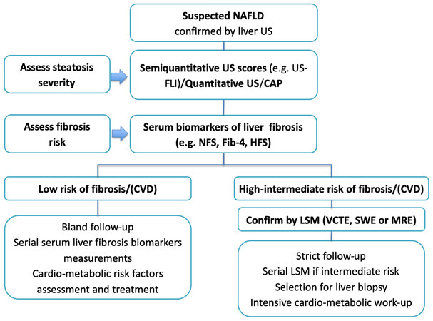
Figure 1. Algorithm to manage NAFLD based on the stratification of the risk of liver-related and CVD complications. The algorithm integrates noninvasive serum biomarkers of liver fibrosis (e.g., NFS, FIB-4, HFS) and sonoelastographic techniques (VCTE, SWE, or MRE) to stratify the risk fibrosis and, as suggested by recent data, the risk of CVD to manage the treatment and follow-up of NAFLD. CAP: Controlled attenuation parameter; CVD: cardiovascular disease; FIB-4: fibrosis-4; HFS: hepamet fibrosis score; LSM: liver stiffness measurement; MRE: magnetic resonance elastography; NAFLD, nonalcoholic fatty liver disease; NFS: NAFLD fibrosis score; SWE: point shear wave elastography; US: ultrasonography; US-FLI: ultrasonographic fatty liver indicator; VCTE: vibration-controlled transient elastography.
Current NAFLD guidelines suggest VCTE and MRE (but not SWE techniques) for LSM because no NAFLD follow-up studies have used SWE; moreover, VCTE and MRE can evaluate liver fibrosis and steatosis quantitively[80].
As reported above, several lines of evidence suggest that more advanced liver fibrosis assessed by noninvasive biomarkers and liver stiffness is associated with liver-related and (especially in T2D patients) CVD outcomes[95-101,104-106]. Therefore, in clinical practice, the combination of various noninvasive markers of NAFLD severity (ultrasonographic steatosis extent, serum biomarkers of fibrosis, and LSM by sono-elastography) permit a noninvasive “one-shot” assessment of hepatic health and CVR among NAFLD individuals. However, longitudinal studies with appropriate follow-up evaluating the association between significant CVEs and suitable evaluation over time to identify MACEs and liver fibrosis biomarkers among NAFLD individuals with/without diabetes are needed to support this practice.
PATHOMECHANISMS AND CLINICAL IMPLICATIONS
Pathophysiology
The pathophysiology associated with the most advanced fibrosing NAFLD/NASH forms with increased risk of atherogenesis is a critical research question and has recently been reviewed in detail[118].
On the one hand, pre-existing NAFLD substantially contributes to incident MetS and its features, such as arterial hypertension and T2D; on the other hand, the most advanced NAFLD/NASH fibrosing forms appear to be specifically associated with MACE. MetS and its components might accelerate macrovascular damage through subclinical systemic inflammation, increased oxidative stress, unbalanced coagulation-fibrinolysis, chronic intermittent hypoxia, hyperuricemia, CKD, pro-inflammatory adipokine profile, excess circulating free fatty acids, and risky lipidomic features[118]. While tending to underestimate the specific pathogenic role of NAFLD, this theory highlights the common involvement of MetS and NAFLD in accelerating atherogenesis.
Other views maintain that the liver is not an innocent bystander and that fibrosing NASH is directly linked with the development of CVD events through increased intrahepatic synthesis of biological mediators such as prothrombogenic factors, fetuin-A, and specific lipidomic profiles typical of unstable coronary plaques[118]. This theory is illustrated in Figure 2 (reprinted, with permission)[118].

Figure 2. “A tale of two pathways”. It is hypothesized that a “long pathway” (blue arrows) leads from steatosis (NAFL) to CVD events through subclinical atherosclerotic burden associated with traditional CVD risk factors. In contrast, a “short pathway” (black arrows) could more directly associate fibrosing NASH with CVD events, possibly through multiple hepatic prothrombogenic factors, fetuin-A, and specific lipidomic signatures. (reprinted, with permission). CVD: Cardiovascular disease; HIF: hypoxia-inducible factor; ICAM-1: intracellular adhesion molecule-1; NAFLD: nonalcoholic fatty liver disease; NASH: nonalcoholic steatohepatitis; VCAM: vascular adhesion molecule-1; VEGF: vascular endothelial growth factor.
Clinical implications
The studies in the present review sustain the theory that noninvasive assessment of hepatic fibrosis can be successfully achieved with algorithms based on anthropometry and laboratory parameters; and with elastometry techniques based on either ultrasonography or MR. The next step could be to combine these techniques in a two-step strategy. To this end, Tamaki et al.[119] conducted a multicenter study of 806 biopsy-proven NAFLD patients to ascertain the accuracy of a diagnosis based on a two-step procedure featuring FIB-4 and MRE. First, individuals exhibiting FIB-4 lower than 1.3 were considered negative irrespective of MRE. Next, if FIB-4 was equal/above 1.3, patients were defined as negative when MRE was lower than 3.6 and positive if MRE was equal/more than 3.6 kPa. The study aimed to compare the diagnostic accuracy of a two-step strategy to MRE alone. The authors found that the AUROC of MRE alone and the two-step strategy were 0.840 and 0.853 in the training cohort (P = 0.4) and 0.867 and 0.834 in the validation cohort
In this evolving scenario, it is becoming increasingly apparent that noninvasive assessment of fibrosis might serve a dual purpose in various clinical settings, notably primary care. On the one hand, it may better stratify those at high risk of fibrosing liver disease; on the other hand, it may simultaneously allow the identification of increased CVR. This finding is critical for personalized and precision medicine approaches that are eagerly awaited for metabolic disorders and NAFLD[120-123]. For example, personalized diagnostic and follow-up schedules might be envisaged for these individuals based on noninvasive fibrosis scores. Importantly, these patients may simultaneously be offered innovative anti-fibrotic agents[124] or drugs targeting hepatic fibrosis and atherogenesis[125].
CONCLUSIONS
While the pathomechanisms associating hepatic fibrosis with CVR and cardiovascular outcomes remain under investigation, accumulating epidemiological and clinical data strongly support that liver fibrosis assessed noninvasively is a risk factor for MACE and mortality. This notion can potentially revolutionize clinical practice in the NAFLD arena and CVR assessment. Further investigation is required to focus on the therapeutic implication of “killing two birds with one stone” using anti-fibrotic agents that combat atherogenesis.
DECLARATIONS
Authors’ contributions
Made substantial contributions to conception and design of the study, wrote the first draft of the manuscript and performed data analysis and interpretation: Ballestri S, Lonardo A
Performed data acquisition and provided administrative, technical, and material support: Di Girolamo M, Baldelli E, Capitelli M
Performed study revision and editing: Ballestri S, Mantovani A, Lonardo A
Availability of data and materials
Not applicable.
Financial support and sponsorship
None.
Conflicts of interest
All authors declared that there are no conflicts of interest.
Ethical approval and consent to participate
Not applicable.
Consent for publication
Not applicable.
Copyright
© The Author(s) 2023.
REFERENCES
1. Parola M, Pinzani M. Liver fibrosis: Pathophysiology, pathogenetic targets and clinical issues. Mol Aspects Med 2019;65:37-55.
2. Lambrecht J, van Grunsven LA, Tacke F. Current and emerging pharmacotherapeutic interventions for the treatment of liver fibrosis. Expert Opin Pharmacother 2020;21:1637-50.
3. Friedman SL, Pinzani M. Hepatic fibrosis 2022: Unmet needs and a blueprint for the future. Hepatology 2022;75:473-88.
4. Gordon SC, Kachru N, Parker E, Korrer S, Ozbay AB, Wong RJ. Health care use and costs among patients with nonalcoholic steatohepatitis with advanced fibrosis using the fibrosis-4 score. Hepatol Commun 2020;4:998-1011.
5. Tacke F, Weiskirchen R. Non-alcoholic fatty liver disease (NAFLD)/non-alcoholic steatohepatitis (NASH)-related liver fibrosis: mechanisms, treatment and prevention. Ann Transl Med 2021;9:729.
6. Friedman SL, Neuschwander-Tetri BA, Rinella M, Sanyal AJ. Mechanisms of NAFLD development and therapeutic strategies. Nat Med 2018;24:908-22.
8. Dulai PS, Singh S, Patel J, et al. Increased risk of mortality by fibrosis stage in nonalcoholic fatty liver disease: systematic review and meta-analysis. Hepatology 2017;65:1557-65.
9. Sanyal AJ, Van Natta ML, Clark J, et al. Prospective study of outcomes in adults with nonalcoholic fatty liver disease. N Engl J Med 2021;385:1559-69.
10. Loomba R, Friedman SL, Shulman GI. Mechanisms and disease consequences of nonalcoholic fatty liver disease. Cell 2021;184:2537-64.
11. Vilar-Gomez E, Calzadilla-Bertot L, Wai-Sun Wong V, et al. Fibrosis severity as a determinant of cause-specific mortality in patients with advanced nonalcoholic fatty liver disease: a multi-national cohort study. Gastroenterology 2018;155:443-457.e17.
12. Mantovani A, Csermely A, Petracca G, et al. Non-alcoholic fatty liver disease and risk of fatal and non-fatal cardiovascular events: an updated systematic review and meta-analysis. Lancet Gastroenterol Hepatol 2021;6:903-13.
13. Sumida Y, Nakajima A, Itoh Y. Limitations of liver biopsy and non-invasive diagnostic tests for the diagnosis of nonalcoholic fatty liver disease/nonalcoholic steatohepatitis. World J Gastroenterol 2014;20:475-85.
14. Lonardo A, Ballestri S. Perspectives of nonalcoholic fatty liver disease research: a personal point of view. Explor Med 2020;1:85-107.
15. Wang FD, Zhou J, Chen EQ. Molecular mechanisms and potential new therapeutic drugs for liver fibrosis. Front Pharmacol 2022;13:787748.
16. Qu W, Ma T, Cai J, et al. Liver fibrosis and MAFLD: from molecular aspects to novel pharmacological strategies. Front Med (Lausanne) 2021;8:761538.
17. Bitto N, Liguori E, La Mura V. Coagulation, microenvironment and liver fibrosis. Cells 2018;7:85.
18. Ballestri S, Capitelli M, Fontana MC, et al. Direct oral anticoagulants in patients with liver disease in the era of non-alcoholic fatty liver disease global epidemic: a narrative review. Adv Ther 2020;37:1910-32.
19. Davis JPE, Caldwell SH. Healing gone wrong: convergence of hemostatic pathways and liver fibrosis? Clin Sci (Lond) 2020;134:2189-201.
20. Mahmoud NI, Messiha BAS, Salehc IG, Abo-Saif AA, Abdel-Bakky MS. Interruption of platelets and thrombin function as a new approach against liver fibrosis induced experimentally in rats. Life Sci 2019;231:116522.
21. Czajka P, Przybyłkowski A, Nowak A, et al. Antiplatelet drugs and liver fibrosis. Platelets 2022;33:219-28.
22. Rahman SR, Roper JA, Grove JI, Aithal GP, Pun KT, Bennett AJ. Integrins as a drug target in liver fibrosis. Liver Int 2022;42:507-21.
23. Dwyer BJ, Macmillan MT, Brennan PN, Forbes SJ. Cell therapy for advanced liver diseases: repair or rebuild. J Hepatol 2021;74:185-99.
24. Wang S, Friedman SL. Hepatic fibrosis: a convergent response to liver injury that is reversible. J Hepatol 2020;73:210-1.
25. Aminian A, Al-Kurd A, Wilson R, et al. Association of bariatric surgery with major adverse liver and cardiovascular outcomes in patients with biopsy-proven nonalcoholic steatohepatitis. J Am Med Assoc 2021;326:2031-42.
26. Chew NWS, Ng CH, Muthiah MD, Sanyal AJ. Comprehensive review and updates on holistic approach towards non-alcoholic fatty liver disease management with cardiovascular disease. Curr Atheroscler Rep 2022;24:515-32.
27. Yang YM, Wang Z, Matsuda M, Seki E. Inhibition of hyaluronan synthesis by 4-methylumbelliferone ameliorates non-alcoholic steatohepatitis in choline-deficient L-amino acid-defined diet-induced murine model. Arch Pharm Res 2021;44:230-40.
28. Pritchard MT, McCracken JM. Identifying novel targets for treatment of liver fibrosis: what can we learn from injured tissues which heal without a scar? Curr Drug Targets 2015;16:1332-46].
29. Andreichenko IN, Tsitrina AA, Fokin AV, et al. 4-methylumbelliferone prevents liver fibrosis by affecting hyaluronan deposition, fstl1 expression and cell localization. Int J Mol Sci 2019;20:6301.
30. Weiz G, Molejon MI, Malvicini M, et al. Glycosylated 4-methylumbelliferone as a targeted therapy for hepatocellular carcinoma. Liver Int 2022;42:444-57.
31. Diagnosis of fibrosis and cirrhosis. Liver biopsy is not always necessary. Prescrire Int 2010;19:38.
33. Vilar-Gomez E, Chalasani N. Non-invasive assessment of non-alcoholic fatty liver disease: Clinical prediction rules and blood-based biomarkers. J Hepatol 2018;68:305-15.
34. Boyle M, Tiniakos D, Schattenberg JM, et al. Performance of the PRO-C3 collagen neo-epitope biomarker in non-alcoholic fatty liver disease. JHEP Rep 2019;1:188-98.
35. Anstee QM, Castera L, Loomba R. Impact of non-invasive biomarkers on hepatology practice: past, present and future. J Hepatol 2022;76:1362-78.
36. Williams AL, Hoofnagle JH. Ratio of serum aspartate to alanine aminotransferase in chronic hepatitis relationship to cirrhosis. Gastroenterology 1988;95:734-9.
37. Forns X, Ampurdanès S, Llovet JM, et al. Identification of chronic hepatitis C patients without hepatic fibrosis by a simple predictive model. Hepatology 2002;36:986-92.
38. Wai CT, Greenson JK, Fontana RJ, et al. A simple noninvasive index can predict both significant fibrosis and cirrhosis in patients with chronic hepatitis C. Hepatology 2003;38:518-26.
39. Harrison SA, Oliver D, Arnold HL, Gogia S, Neuschwander-Tetri BA. Development and validation of a simple NAFLD clinical scoring system for identifying patients without advanced disease. Gut 2008;57:1441-7.
40. Sterling RK, Lissen E, Clumeck N, et al. Development of a simple noninvasive index to predict significant fibrosis in patients with HIV/HCV coinfection. Hepatology 2006;43:1317-25.
41. Angulo P, Hui JM, Marchesini G, et al. The NAFLD fibrosis score: a noninvasive system that identifies liver fibrosis in patients with NAFLD. Hepatology 2007;45:846-54.
42. McPherson S, Hardy T, Dufour JF, et al. Age as a confounding factor for the accurate non-invasive diagnosis of advanced NAFLD fibrosis. Am J Gastroenterol 2017;112:740-51.
43. Lemoine M, Shimakawa Y, Nayagam S, et al. The gamma-glutamyl transpeptidase to platelet ratio (GPR) predicts significant liver fibrosis and cirrhosis in patients with chronic HBV infection in West Africa. Gut 2016;65:1369-76.
44. Ampuero J, Pais R, Aller R, et al. HEPAmet Registry. Development and validation of hepamet fibrosis scoring system-a simple, noninvasive test to identify patients with nonalcoholic fatty liver disease with advanced fibrosis. Clin Gastroenterol Hepatol 2020;18:216-225.e5.
45. Ballestri S, Mantovani A, Baldelli E, et al. Liver fibrosis biomarkers accurately exclude advanced fibrosis and are associated with higher cardiovascular risk scores in patients with NAFLD or viral chronic liver disease. Diagnostics (Basel) 2021;11:98.
46. Brandman D, Boyle M, McPherson S, et al. Comparison of clinical prediction rules for ruling out cirrhosis in nonalcoholic fatty liver disease (NAFLD). Aliment Pharmacol Ther 2022;55:1441-51.
47. Sun W, Cui H, Li N, et al. Comparison of FIB-4 index, NAFLD fibrosis score and BARD score for prediction of advanced fibrosis in adult patients with non-alcoholic fatty liver disease: a meta-analysis study. Hepatol Res 2016;46:862-70.
48. Boursier J, Canivet CM, Costentin C, et al. Impact of type 2 diabetes on the accuracy of noninvasive tests of liver fibrosis with resulting clinical implications. Clin Gastroenterol Hepatol ;2022:S1542-3565(22)00248.
49. Petta S, Vanni E, Bugianesi E, et al. The combination of liver stiffness measurement and NAFLD fibrosis score improves the noninvasive diagnostic accuracy for severe liver fibrosis in patients with nonalcoholic fatty liver disease. Liver Int 2015;35:1566-73.
50. Jafarov F, Kaya E, Bakir A, Eren F, Yilmaz Y. The diagnostic utility of fibrosis-4 or nonalcoholic fatty liver disease fibrosis score combined with liver stiffness measurement by fibroscan in assessment of advanced liver fibrosis: a biopsy-proven nonalcoholic fatty liver disease study. Eur J Gastroenterol Hepatol 2020;32:642-9.
51. Newsome PN, Sasso M, Deeks JJ, et al. FibroScan-AST (FAST) score for the non-invasive identification of patients with non-alcoholic steatohepatitis with significant activity and fibrosis: a prospective derivation and global validation study. Lancet Gastroenterol Hepatol 2020;5:362-73.
52. Eddowes PJ, Sasso M, Allison M, et al. Accuracy of fibroscan controlled attenuation parameter and liver stiffness measurement in assessing steatosis and fibrosis in patients with nonalcoholic fatty liver disease. Gastroenterology 2019;156:1717-30.
53. Oeda S, Takahashi H, Imajo K, et al. Diagnostic accuracy of FibroScan-AST score to identify non-alcoholic steatohepatitis with significant activity and fibrosis in Japanese patients with non-alcoholic fatty liver disease: comparison between M and XL probes. Hepatol Res 2020;50:831-9.
54. Kim BK, Tamaki N, Imajo K, et al. Head-to-head comparison between MEFIB, MAST, and FAST for detecting stage 2 fibrosis or higher among patients with NAFLD. J Hepatol 2022;77:1482-90.
55. Ng CH, Lim WH, Hui Lim GE, et al. Mortality outcomes by fibrosis stage in nonalcoholic fatty liver disease: a systematic review and meta-analysis. Clin Gastroenterol Hepatol ;2022:S1542-3565(22)00439.
56. Reddy YK, Marella HK, Jiang Y, et al. Natural history of non-alcoholic fatty liver disease: a study with paired liver biopsies. J Clin Exp Hepatol 2020;10:245-54.
57. Perazzo H, Munteanu M, Ngo Y, et al. Prognostic value of liver fibrosis and steatosis biomarkers in type-2 diabetes and dyslipidaemia. Aliment Pharmacol Ther 2014;40:1081-93.
58. Kim D, Kim WR, Kim HJ, Therneau TM. Association between noninvasive fibrosis markers and mortality among adults with nonalcoholic fatty liver disease in the United States. Hepatology 2013;57:1357-65.
59. Song DS, Chang UI, Kang SG, Song SW, Yang JM. Noninvasive serum fibrosis markers are associated with coronary artery calcification in patients with nonalcoholic fatty liver disease. Gut Liver 2019;13:658-68.
60. Schonmann Y, Yeshua H, Bentov I, Zelber-Sagi S. Liver fibrosis marker is an independent predictor of cardiovascular morbidity and mortality in the general population. Dig Liver Dis 2021;53:79-85.
61. Han E, Lee YH, Lee JS, et al. Fibrotic burden determines cardiovascular risk among subjects with metabolic dysfunction-associated fatty liver disease. Gut Liver 2022;16:786-97.
62. Chen Q, Li Q, Li D, et al. Association between liver fibrosis scores and the risk of mortality among patients with coronary artery disease. Atherosclerosis 2020;299:45-52.
63. Baratta F, Pastori D, Angelico F, et al. Nonalcoholic fatty liver disease and fibrosis associated with increased risk of cardiovascular events in a prospective study. Clin Gastroenterol Hepatol 2020;18:2324-2331.e4.
64. Lee J, Kim HS, Cho YK, et al. Association between noninvasive assessment of liver fibrosis and coronary artery calcification progression in patients with nonalcoholic fatty liver disease. Sci Rep 2020;10:18323.
65. Liu HH, Cao YX, Jin JL, et al. Liver fibrosis scoring systems as novel tools for predicting cardiovascular outcomes in patients following elective percutaneous coronary intervention. J Am Heart Assoc 2021;10:e018869.
66. Peters AE, Pandey A, Ayers C, et al. Association of liver fibrosis risk scores with clinical outcomes in patients with heart failure with preserved ejection fraction: findings from TOPCAT. ESC Heart Fail 2021;8:842-8.
67. Parikh NS, Koh I, VanWagner LB, Elkind MSV, Zakai NA, Cushman M. Liver fibrosis is associated with ischemic stroke risk in women but not men: the REGARDS study. J Stroke Cerebrovasc Dis 2021;30:105788.
68. Oh TJ, Kim K, Moon JH, Choi SH, Cho NH, Jang HC. Liver fibrosis indices for the prediction of mortality in korean subjects: a 16-year prospective cohort study. J Endocr Soc 2021;5:bvab123.
69. Delgado GE, Kleber ME, Moissl AP, et al. Surrogate scores of advanced fibrosis in NAFLD/NASH do not predict mortality in patients with medium-to-high cardiovascular risk. Am J Physiol Gastrointest Liver Physiol 2021;321:G252-61.
70. Tamaki N, Kurosaki M, Takahashi Y, et al. Liver fibrosis and fatty liver as independent risk factors for cardiovascular disease. J Gastroenterol Hepatol 2021;36:2960-6.
71. Akuta N, Kawamura Y, Arase Y, et al. PNPLA3 genotype and fibrosis-4 index predict cardiovascular diseases of Japanese patients with histopathologically-confirmed NAFLD. BMC Gastroenterol 2021;21:434.
72. Jin JL, Zhang HW, Cao YX, et al. Liver fibrosis scores and coronary atherosclerosis: novel findings in patients with stable coronary artery disease. Hepatol Int 2021;15:413-23.
73. Zupo R, Castellana F, De Nucci S, et al. Liver fibrosis and 8-year all-cause mortality trajectories in the aging cohort of the salus in apulia study. Biomedicines 2021;9:1617.
74. Zupo R, Castellana F, Donghia R, et al. Liver frailty and all-cause mortality in the older participants of the Salus in Apulia study. Geroscience 2022;44:835-45.
75. Vieira Barbosa J, Milligan S, Frick A, et al. Fibrosis-4 index can independently predict major adverse cardiovascular events in nonalcoholic fatty liver disease. Am J Gastroenterol 2022;117:453-61.
76. Ferraioli G, Wong VW, Castera L, et al. Liver ultrasound elastography: an update to the world federation for ultrasound in medicine and biology guidelines and recommendations. Ultrasound Med Biol 2018;44:2419-40.
77. Ballestri S, Nascimbeni F, Lugari S, Lonardo A, Francica G. A critical appraisal of the use of ultrasound in hepatic steatosis. Expert Rev Gastroenterol Hepatol 2019;13:667-81.
79. Lee MS, Bae JM, Joo SK, Woo H, Lee DH, Jung YJ, et al. Prospective comparison among transient elastography, supersonic shear imaging, and ARFI imaging for predicting fibrosis in nonalcoholic fatty liver disease. PLoS One 2017;12:e0188321.
80. Honda Y, Yoneda M, Imajo K, Nakajima A. Elastography techniques for the assessment of liver fibrosis in non-alcoholic fatty liver disease. Int J Mol Sci 2020;21:4039.
81. Conti CB, Cavalcoli F, Fraquelli M, Conte D, Massironi S. Ultrasound elastographic techniques in focal liver lesions. World J Gastroenterol 2016;22:2647-56.
82. Liu H, Fu J, Hong R, Liu L, Li F. Acoustic radiation force impulse elastography for the non-invasive evaluation of hepatic fibrosis in non-alcoholic fatty liver disease patients: a systematic review & meta-analysis. PLoS One 2015;10:e0127782.
83. Cassinotto C, Boursier J, de Lédinghen V, et al. Liver stiffness in nonalcoholic fatty liver disease: a comparison of supersonic shear imaging, FibroScan, and ARFI with liver biopsy. Hepatology 2016;63:1817-27.
84. Jiang W, Huang S, Teng H, et al. Diagnostic accuracy of point shear wave elastography and transient elastography for staging hepatic fibrosis in patients with non-alcoholic fatty liver disease: a meta-analysis. BMJ Open 2018;8:e021787.
85. Herrmann E, de Lédinghen V, Cassinotto C, et al. Assessment of biopsy-proven liver fibrosis by two-dimensional shear wave elastography: an individual patient data-based meta-analysis. Hepatology 2018;67:260-72.
86. Castera L, Friedrich-Rust M, Loomba R. Noninvasive assessment of liver disease in patients with nonalcoholic fatty liver disease. Gastroenterology 2019;156:1264-1281.e4.
87. Selvaraj EA, Mózes FE, Jayaswal ANA, et al. Diagnostic accuracy of elastography and magnetic resonance imaging in patients with NAFLD: a systematic review and meta-analysis. J Hepatol 2021;75:770-85.
88. Petta S, Sebastiani G, Bugianesi E, et al. Non-invasive prediction of esophageal varices by stiffness and platelet in non-alcoholic fatty liver disease cirrhosis. J Hepatol 2018;69:878-85.
89. Berzigotti A, Ferraioli G, Bota S, Gilja OH, Dietrich CF. Novel ultrasound-based methods to assess liver disease: the game has just begun. Dig Liver Dis 2018;50:107-12.
90. Park CC, Nguyen P, Hernandez C, et al. Magnetic resonance elastography vs transient elastography in detection of fibrosis and noninvasive measurement of steatosis in patients with biopsy-proven nonalcoholic fatty liver disease. Gastroenterology 2017;152:598-607.e2.
91. Lee YS, Lee JE, Yi HS, et al. MRE-based NASH score for diagnosis of nonalcoholic steatohepatitis in patients with nonalcoholic fatty liver disease. Hepatol Int 2022;16:316-24.
92. Polyzos SA, Kechagias S, Tsochatzis EA. Review article: non-alcoholic fatty liver disease and cardiovascular diseases: associations and treatment considerations. Aliment Pharmacol Ther 2021;54:1013-25.
93. Veracruz N, Hameed B, Saab S, Wong RJ. The association between nonalcoholic fatty liver disease and risk of cardiovascular disease, stroke, and extrahepatic cancers. J Clin Exp Hepatol 2021;11:45-81.
94. Friedrich-Rust M, Schoelzel F, Maier S, et al. Severity of coronary artery disease is associated with non-alcoholic fatty liver dis-ease: a single-blinded prospective mono-center study. PLoS One 2017;12:e0186720.
95. Song Y, Dang Y, Wang P, Tian G, Ruan L. CHD is associated with higher grades of NAFLD predicted by liver stiffness. J Clin Gastroenterol 2020;54:271-7.
96. Lombardi R, Airaghi L, Targher G, et al. Liver fibrosis by FibroScan® independently of established cardiovascular risk parameters associates with macrovascular and microvascular complications in patients with type 2 diabetes. Liver Int 2020;40:347-54.
97. Mantovani A, Turino T, Lando MG, et al. Screening for non-alcoholic fatty liver disease using liver stiffness measurement and its association with chronic kidney disease and cardiovascular complications in patients with type 2 diabetes. Diabetes Metab 2020;46:296-303.
98. Mikolasevic I, Rahelic D, Turk-Wensween T, et al. Significant liver fibrosis, as assessed by fibroscan, is independently associated with chronic vascular complications of type 2 diabetes: a multicenter study. Diabetes Res Clin Pract 2021;177:108884.
99. Long MT, Zhang X, Xu H, et al. Hepatic fibrosis associates with multiple cardiometabolic disease risk factors: the framingham heart study. Hepatology 2021;73:548-59.
100. Pennisi G, Di Marco V, Buscemi C, et al. Interplay between non-alcoholic fatty liver disease and cardiovascular risk in an asymptomatic general population. J Gastroenterol Hepatol 2021;36:2389-96.
101. Park JG, Jung J, Verma KK, et al. Liver stiffness by magnetic resonance elastography is associated with increased risk of cardiovascular disease in patients with non-alcoholic fatty liver disease. Aliment Pharmacol Ther 2021;53:1030-7.
102. Ciardullo S, Cannistraci R, Mazzetti S, Mortara A, Perseghin G. Nonalcoholic fatty liver disease, liver fibrosis and cardiovascular disease in the adult US population. Front Endocrinol 2021;12:711484.
103. Liu K, Wong VW, Lau K, et al. Prognostic value of controlled attenuation parameter by transient elastography. Am J Gastroenterol 2017;112:1812-23.
104. Shili-Masmoudi S, Wong GL, Hiriart JB, et al. Liver stiffness measurement predicts long-term survival and complications in non-alcoholic fatty liver disease. Liver Int 2020;40:581-9.
105. Mikolasevic I, Domislovic V, Ruzic A, et al. Elastographic parameters of liver steatosis and fibrosis predict independently the risk of incident chronic kidney disease and acute myocardial infarction in patients with type 2 diabetes mellitus. J Diabetes Complicat 2022;36:108226.
106. Cardoso CRL, Villela-Nogueira CA, Leite NC, Salles GF. Prognostic impact of liver fibrosis and steatosis by transient elastography for cardiovascular and mortality outcomes in individuals with nonalcoholic fatty liver disease and type 2 diabetes: the Rio de Janeiro Cohort study. Cardiovasc Diabetol 2021;20:193.
107. Grgurevic I, Salkic N, Mustapic S, et al. Liver and nonliver-related outcomes at 2 years are not influenced by the results of the fib-4 test and liver elastography in a real-life cohort of patients with type 2 diabetes. Can J Gastroenterol Hepatol 2021;2021:5582813.
108. Petta S, Sebastiani G, Viganò M, et al. Monitoring occurrence of liver-related events and survival by transient elastography in patients with nonalcoholic fatty liver disease and compensated advanced chronic liver disease. Clin Gastroenterol Hepatol 2021;19:806-815.e5.
109. Mangla N, Ajmera VH, Caussy C, et al. Liver stiffness severity is associated with increased cardiovascular risk in patients with type 2 diabetes. Clin Gastroenterol Hepatol 2020;18:744-746.e1.
110. Johnson AL, Hayward KL, Patel P, et al. Predicting liver-related outcomes in people with nonalcoholic fatty liver disease: the prognostic value of noninvasive fibrosis tests. Hepatol Commun 2022;6:728-39.
111. Petta S, Wong VW, Cammà C, et al. Serial combination of non-invasive tools improves the diagnostic accuracy of severe liver fibrosis in patients with NAFLD. Aliment Pharmacol Ther 2017;46:617-27.
112. Chan WK, Treeprasertsuk S, Goh GB, et al. Optimizing use of nonalcoholic fatty liver disease fibrosis score, fibrosis-4 score, and liver stiffness measurement to identify patients with advanced fibrosis. Clin Gastroenterol Hepatol 2019;17:2570-2580.e37.
113. Ballestri S, Romagnoli D, Nascimbeni F, Francica G, Lonardo A. Role of ultrasound in the diagnosis and treatment of nonalcoholic fatty liver disease and its complications. Expert Rev Gastroenterol Hepatol 2015;9:603-27.
114. Targher G, Byrne CD, Lonardo A, Zoppini G, Barbui C. Non-alcoholic fatty liver disease and risk of incident cardiovascular disease: a meta-analysis. J Hepatol 2016;65:589-600.
115. Ballestri S, Mantovani A, Nascimbeni F, Lugari S, Lonardo A. Extra-hepatic manifestations and complications of nonalcoholic fatty liver disease. Future Med Chem 2019;11:2171-92.
116. Ballestri S, Tana C, Di Girolamo M, et al. Semi-quantitative ultrasonographic evaluation of NAFLD. Curr Pharm Des 2020;26:3915-27.
117. Nascimbeni F, Ballestri S, Machado MV, et al. Clinical relevance of liver histopathology and different histological classifications of NASH in adults. Expert Rev Gastroenterol Hepatol 2018;12:351-67.
118. Lonardo A, Nascimbeni F, Mantovani A, Targher G. Hypertension, diabetes, atherosclerosis and NASH: cause or consequence? J Hepatol 2018;68:335-52.
119. Tamaki N, Imajo K, Sharpton SR, et al. Two-step strategy, fib-4 followed by magnetic resonance elastography, for detecting advanced fibrosis in NAFLD. Clin Gastroenterol Hepatol ;2022:S1542-3565(22)00077.
121. Lonardo A, Byrne CD, Targher G. Precision medicine approaches in metabolic disorders and target organ damage: where are we now, and where are we going? Metab Target Organ Damage 2021; doi: 10.20517/mtod.2021.03.
121. Lonardo A. Renaming NAFLD to MAFLD: could the LDE system assist in this transition? J Clin Med 2021;10:492.
122. Lonardo A, Arab JP, Arrese M. Perspectives on precision medicine approaches to nafld diagnosis and management. Adv Ther 2021;38:2130-58.
123. Lonardo A. Precision medicine in nonalcoholic fatty liver disease. J Gastroenterol Hepatol 2022;37:1175-8.
124. Alkhouri N, Herring R, Kabler H, et al. Safety and efficacy of combination therapy with semaglutide, cilofexor and firsocostat in patients with non-alcoholic steatohepatitis: a randomised, open-label phase II trial. J Hepatol 2022;77:607-18.
Cite This Article
Export citation file: BibTeX | RIS
OAE Style
Ballestri S, Mantovani A, Di Girolamo M, Baldelli E, Capitelli M, Lonardo A. Liver fibrosis in nonalcoholic fatty liver disease patients: noninvasive evaluation and correlation with cardiovascular disease and mortality. Metab Target Organ Damage 2023;3:1. http://dx.doi.org/10.20517/mtod.2022.23
AMA Style
Ballestri S, Mantovani A, Di Girolamo M, Baldelli E, Capitelli M, Lonardo A. Liver fibrosis in nonalcoholic fatty liver disease patients: noninvasive evaluation and correlation with cardiovascular disease and mortality. Metabolism and Target Organ Damage. 2023; 3(1): 1. http://dx.doi.org/10.20517/mtod.2022.23
Chicago/Turabian Style
Ballestri, Stefano, Alessandro Mantovani, Maria Di Girolamo, Enrica Baldelli, Mariano Capitelli, Amedeo Lonardo. 2023. "Liver fibrosis in nonalcoholic fatty liver disease patients: noninvasive evaluation and correlation with cardiovascular disease and mortality" Metabolism and Target Organ Damage. 3, no.1: 1. http://dx.doi.org/10.20517/mtod.2022.23
ACS Style
Ballestri, S.; Mantovani A.; Di Girolamo M.; Baldelli E.; Capitelli M.; Lonardo A. Liver fibrosis in nonalcoholic fatty liver disease patients: noninvasive evaluation and correlation with cardiovascular disease and mortality. Metab Target Organ Damage. 2023, 3, 1. http://dx.doi.org/10.20517/mtod.2022.23
About This Article
Special Issue
Copyright

Data & Comments
Data


 Cite This Article 58 clicks

 Like This Article 19
likes
Like This Article 19
likes


Comments
Comments must be written in English. Spam, offensive content, impersonation, and private information will not be permitted. If any comment is reported and identified as inappropriate content by OAE staff, the comment will be removed without notice. If you have any queries or need any help, please contact us at support@oaepublish.com.